

 , Şükriye Ebru Önder 1
, Serdar Bozyel 1
, Şadiye Nur Dalgıç 1
, Osman Muhsin Çelik 1
, Metin Çağdaş 1
, Tümer Erdem Güler 1
, Müjdat Aktaş 2
, Tolga Aksu 3
, Şükriye Ebru Önder 1
, Serdar Bozyel 1
, Şadiye Nur Dalgıç 1
, Osman Muhsin Çelik 1
, Metin Çağdaş 1
, Tümer Erdem Güler 1
, Müjdat Aktaş 2
, Tolga Aksu 3
2Department of Cardiology, Health Sciences University, Prof. Dr. Cemil Taşçıoğlu City Hospital, İstanbul, Türkiye
3Department of Cardiology, İstanbul Aydın University, Medical Park Florya Hospital, İstanbul, Türkiye
Abstract
Background: Cardiac resynchronization therapy (CRT) is an established treatment for selected patients with heart failure (HF); however, a substantial proportion of patients do not derive clinical or echocardiographic benefit. Systemic inflammation has been implicated in adverse myocardial remodeling and may influence response to CRT. This study aimed to evaluate the association between the systemic immune-inflammation index (SII) and CRT response.
Methods: This retrospective, single-center study included 110 patients with HF who underwent CRT implantation. Clinical, echocardiographic, and laboratory parameters, including SII, were assessed at baseline and at 6-month follow-up. Cardiac resynchronization therapy response was defined using combined echocardiographic and clinical criteria. Associations between inflammatory markers and CRT response were analyzed.
Results: Nonresponders demonstrated significantly higher baseline SII levels compared with responders. During follow-up, SII values increased significantly in nonresponders, whereas no significant change was observed in responders. Baseline C-reactive protein (CRP) levels were similar between groups. Chronic ischemic heart disease and elevated baseline SII were independently associated with CRT nonresponse. The discriminatory ability of baseline SII for CRT nonresponse was moderate.
Conclusion: Elevated SII was associated with an increased likelihood of CRT nonresponse and persistent inflammatory activity during follow-up. These findings suggest that SII reflects an unfavorable inflammatory and biological milieu related to impaired CRT response, rather than a direct causal mechanism. Systemic immune-inflammation index may serve as a complementary biomarker for risk stratification in patients undergoing CRT.
Graphical Abstract
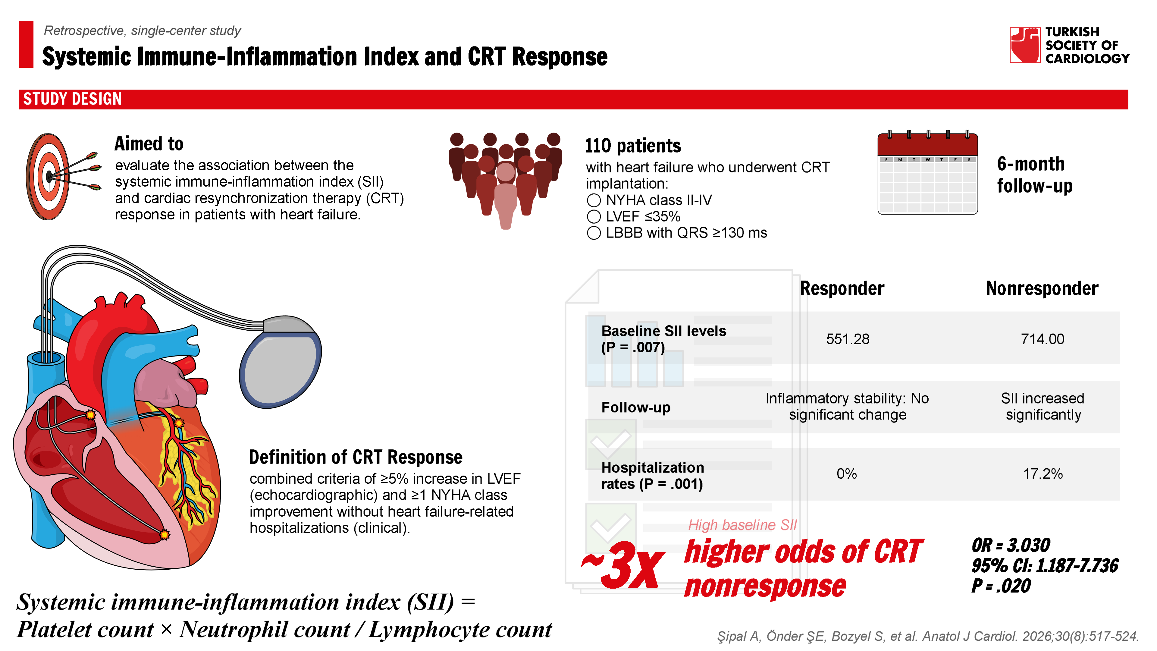
Highlights
- Elevated baseline systemic immune-inflammation index (SII) levels were associated with nonresponse to cardiac resynchronization therapy (CRT).
- During follow-up, SII values increased significantly in nonresponders, whereas responders demonstrated relative inflammatory stability rather than a significant reduction in SII.
- High baseline SII levels were independently associated with a significantly increased likelihood of being a CRT nonresponder and with poorer clinical and echocardiographic outcomes.
- Persistent elevation of inflammatory biomarkers—including SII, C-reactive protein, B-type natriuretic peptide (BNP), and troponin—in nonresponders suggests that ongoing systemic inflammation may limit CRT efficacy.
- These findings indicate that SII may serve as a complementary, easily accessible biomarker for patient characterization and risk stratification before CRT implantation.
Introduction
Heart failure (HF) is a progressive clinical syndrome characterized by impaired cardiac function and substantial morbidity and mortality. Despite advances in pharmacological therapy, many patients remain symptomatic. Cardiac resynchronization therapy (CRT) improves symptoms, ventricular function, and survival in appropriately selected patients
The variability in CRT response has prompted extensive investigation into clinical, electrocardiographic, imaging, and laboratory predictors.
The systemic immune-inflammation index (SII), derived from neutrophil, lymphocyte, and platelet counts, integrates inflammatory, immune, and prothrombotic pathways. Although SII has been evaluated in several cardiovascular conditions, its association with CRT response has not been adequately explored. Therefore, this study aimed to investigate the relationship between SII and CRT response, with particular emphasis on distinguishing association from causation.
Methods
Patients
The study population consisted of 110 HF patients who underwent CRT implantation in the center between January 2019 and January 2024, in accordance with the current guideline recommendations. The study included 112 HF patients who met the following criteria:
Patients were excluded from the study if they had the following:
Additionally, patients with a life expectancy of less than 12 months or a follow-up period shorter than 6 months were not included. This study is a retrospective analysis of prospectively collected data, and institutional ethics approval was obtained in accordance with the principles of the Helsinki Declaration.
Blood Samples
Venous blood samples were collected before the procedure and at the sixth-month follow-up. Laboratory parameters such as hemoglobin, creatinine, leukocytes, platelets, neutrophils, lymphocytes, C-reactive protein (CRP), and low-density lipoprotein (LDL) levels were recorded. Based on these results, the SII was calculated using the formula: SII = Platelet count × Neutrophil count / Lymphocyte count.
Clinical Evaluation
All patients underwent a clinical evaluation, including NYHA classification, QRS duration, and comorbidity assessment. This was performed by an independent physician blinded to other data. QRS duration was measured on surface ECG, taking the widest QRS complex from leads II, V1, and V6. Patients were evaluated for age, sex, history of coronary artery disease, diabetes, hyperlipidemia, hypertension, and other comorbidities.
Patients with a history of coronary artery disease and/or prior myocardial infarction were classified as ischemic. Those without a history of myocardial infarction or revascularization and with no more than 50% atherosclerotic lesions in 2 or more epicardial vessels, the left main coronary artery, or the proximal left anterior descending artery were classified as non-ischemic.
Echocardiography
Standard echocardiographic imaging was performed both before CRT implantation and at the sixth-month follow-up. Echocardiographic imaging was conducted in a resting state with the patient in the left lateral decubitus position, using the equipment available in the clinic (Vivid 7 Ultrasound System; GE, Horten, Norway). Measurements were made according to the recommendations of the American Society of Echocardiography. The LVEF was calculated using the modified Simpson method.
Device Implantation and Optimization Protocol
All patients received a CRT device with a defibrillator (CRT-D). The CRT-D implantation was performed transvenously via the left subclavian route. Before the placement of the left ventricular (LV) lead, routine coronary sinus venography was performed. The LV lead was preferably positioned in the lateral or posterolateral branches of the coronary sinus. The right atrial lead was implanted in the atrial appendage, and the right ventricular lead was positioned in the apex region. Atrioventricular (AV) and interventricular interval optimization was performed by an experienced cardiologist using Doppler echocardiography, with transmitral flow and velocity-time integral measurements used to achieve the shortest possible QRS duration and maximize ventricular synchrony. To evaluate the potential impact of LV lead positioning on CRT response, pacing sites (posterior, posterolateral, and lateral coronary sinus branches) between responders and nonresponders were compared, with lead positioning determined using fluoroscopic guidance during implantation. To further assess its influence on CRT outcomes, a secondary analysis was conducted at the 6-month follow-up, evaluating the relationship between lead position and key echocardiographic and clinical parameters, including LVEF improvement, NYHA class reduction, and hospitalization rates.
Definition of Cardiac Resynchronization Therapy Response
Left ventricular reverse remodeling was assessed quantitatively based on the improvement in LVEF following CRT, with an increase of ≥5% at the 6-month follow-up considered indicative of a positive echocardiographic response. Clinical response was defined as an improvement of at least 1 NYHA functional class level without any HF-related hospitalizations during the follow-up period. Patients meeting both echocardiographic and clinical response criteria were classified as responders, while those who did not fulfill these criteria were classified as nonresponders.
Statistical Analysis
The statistical analyses were conducted using IBM SPSS Statistics v26 software. The normality of the variables was assessed using the Kolmogorov–Smirnov test. The chi-squared test and Fisher’s exact test were utilized to compare categorical variables. Fisher’s exact test was applied when more than 20% of the cells had expected frequencies less than 5, while the chi-squared test was used when this condition was not met.
Ethical Approval
This study was approved by an independent Institutional Ethics Committee (Approval Date: November 13, 2025; Decision Number: 2025-128) and conducted in accordance with the principles of the Declaration of Helsinki. Because this research involved a retrospective analysis of previously recorded clinical data, the requirement for informed consent was waived by the ethics committee.
Results
A total of 110 patients who met the inclusion criteria and experienced no complications were included in the study. Of these, 35.5% (n = 39) were female, and 64.5% (n = 71) were male. Among the participants, 43.6% (n = 48) had chronic ischemic heart disease, 21.1% (n = 23) had diabetes, and 30.9% (n = 34) had hypertension. Pacing was performed in the posterior branch in 72.7% (n = 80), the posterolateral branch in 20.9% (n = 23), and the lateral branch in 6.4% (n = 7). Symptomatically, 45.5% (n = 50) of the patients were classified as NYHA Class II, and 54.5% (n = 60) as Class III prior to the procedure. Echocardiographic evaluations conducted before the procedure showed mild mitral regurgitation in 62.7% (n = 69), moderate mitral regurgitation in 29.1% (n = 32), and severe mitral regurgitation in 8.2% (n = 9).
Patients were divided into 2 groups based on their response to CRT: responders (n = 81) and nonresponders (n = 29). There were no significant differences between the groups in terms of age (
Baseline LVEF was similar between groups, with median values of 28% (nterquartile range [IQR]: 25-32) in responders and 28% (IQR: 25-31) in nonresponders (
The distribution of LV lead placement sites was comparable between CRT responders and nonresponders. Posterior, posterolateral, and lateral pacing sites showed no significant differences between groups (
Analysis of changes in SII between baseline and follow-up showed a statistically significant increase in SII values in the nonresponder group (
At the 6-month follow-up, significant differences were observed between the groups in hematological, biochemical, and echocardiographic parameters (
Regarding clinical outcomes, hospitalization rates were significantly higher in the nonresponder group (
Logistic regression analysis identified chronic ischemic heart disease and high baseline SII levels as significant predictors of CRT nonresponse. The model achieved an overall accuracy of 74.5%, correctly classifying 44.8% of nonresponders and 85.2% of responders. Chronic ischemic heart disease was associated with a higher likelihood of nonresponse (
The AUC for the ROC analysis of the baseline SII index was 0.669 (Figure 1), with a standard error of 0.059 and a
At 6 months, no significant association was observed between LV lead positioning and clinical or echocardiographic response to CRT. Patients with posterior, posterolateral, and lateral lead placements exhibited similar rates of LVEF improvement and NYHA class reduction (
Baseline characteristics were comparable between responders and nonresponders with respect to age, sex, and major comorbidities, except for a higher prevalence of chronic ischemic heart disease among nonresponders. Baseline SII levels were significantly higher in nonresponders, whereas baseline CRP levels did not differ between groups.
During follow-up, SII values increased significantly in the nonresponder group, indicating persistent or worsening systemic inflammation. In contrast, no significant difference was observed between baseline and follow-up SII values in the responder group, suggesting relative inflammatory stability.
QRS duration at follow-up was longer in nonresponders. Given the standardized implantation and optimization protocol, this finding was interpreted as reflecting differences in electrical remodeling and myocardial substrate rather than unequal CRT optimization.
In the nonresponder group, 5 patients (17%) experienced HF-related hospitalization during the follow-up period. Importantly, all baseline blood samples were obtained during clinically stable conditions and at least 4 weeks after the last episode of hospitalization or acute decompensation. Patients with blood sampling performed during acute decompensated HF were not included in the analysis.
Discussion
The principal finding of this study is that elevated SII is associated with CRT nonresponse and with persistent systemic inflammation during follow-up. Importantly, these findings should be interpreted as associative rather than causal. At baseline, higher SII levels in nonresponders (
Cardiac resynchronization therapy is an effective treatment modality for patients with HF and prolonged QRS duration, aimed at improving symptoms, enhancing quality of life, and reducing mortality risk.
Systemic immune-inflammation index has emerged in recent years as a valuable biomarker that reflects a combination of inflammatory and thrombotic processes in various clinical contexts. Calculated using neutrophil, platelet, and lymphocyte counts, SII serves as a comprehensive measure of inflammatory burden and immune response.17
In this study, the aim was to investigate the role of the SII as a prognostic marker in HF patients undergoing CRT. Given the potential impact of systemic inflammation on CRT outcomes, previous research has explored its influence on patient response rates and clinical prognoses. Stavrakis et al17 previously reported that elevated inflammatory markers negatively impact response rates and are associated with worse clinical outcomes in CRT patients. Similarly, high SII values have been shown to reflect increased inflammatory burden and thrombotic activity, which may limit the effectiveness of CRT.
In the present study, responders did not demonstrate a significant change in SII values between baseline and follow-up, indicating relative inflammatory stability over time. In contrast, nonresponders exhibited a significant increase in SII levels, reflecting persistent or worsening systemic inflammation. This divergence suggests that the absence of inflammatory progression, rather than a reduction in inflammatory burden, may characterize patients who respond favorably to CRT. Within this context, previous studies have suggested that favorable ventricular remodeling after CRT may be accompanied by attenuation of systemic inflammatory activity, particularly in selected patient populations. Karaüzüm et al19 demonstrated that a reduction in inflammation following CRT is associated with improved LVEF levels. This suggests that CRT not only provides mechanical hemodynamic benefits but also supports myocardial recovery by reducing inflammation. This effect was particularly pronounced in patients with low baseline SII values.
The NYHA classification is a standard tool for assessing the severity of HF symptoms. In this study, higher baseline SII values were associated with more severe NYHA classes (III-IV), and the responder group exhibited improvements in NYHA classification alongside reductions in SII following CRT. Specifically, patients in NYHA III-IV who experienced symptomatic improvement after CRT demonstrated significant decreases in SII, underscoring the role of inflammation in CRT success. Cleland et al15 previously reported that CRT supports symptomatic improvement, particularly in patients with NYHA III-IV classification. Given that SII reflects both inflammatory and thrombotic processes, it may serve as a more comprehensive predictor of NYHA class and CRT response.
Mitral regurgitation (MR) is a common complication in HF patients and has the potential to improve through the hemodynamic benefits of CRT. In this study, higher baseline SII values were associated with more severe MR, and significant improvement in MR severity was observed in the responder group alongside reductions in SII following CRT. Di Biase et al21 reported similar findings, demonstrating significant MR improvement in patients who exhibited reduced inflammatory markers post-CRT. This suggests that CRT mitigates the burden on mitral valve dysfunction by modulating inflammation, thereby supporting MR improvement.
The relationship between baseline and 6-month SII values and QRS durations provides valuable insights into the electrical effects of CRT. Elevated pre-CRT SII values were generally associated with prolonged QRS durations, and patients with higher baseline SII values showed limited QRS shortening following CRT. In contrast, the responder group exhibited significant QRS shortening, accompanied by reductions in SII. The potential pathophysiological mechanisms underlying these findings, as noted in previous studies, include the role of heightened inflammation in promoting myocardial fibrosis, which impairs ventricular synchronization and prolongs QRS duration.
To comprehensively evaluate inflammation’s impact on CRT response, this study assessed hematological and biochemical markers such as CRP, neutrophils, platelets, neutrophil-to-lymphocyte ratio (NLR), and platelet-to-lymphocyte ratio (PLR). Elevated levels of these parameters in nonresponders indicate persistent inflammation and associated thrombotic activity, potentially impairing ventricular remodeling and limiting CRT efficacy. Conversely, responders demonstrated significantly lower and decreasing levels of these inflammatory markers post-CRT, highlighting the role of inflammation control in enhancing CRT’s mechanical and clinical benefits.
Previous investigations have shown that long-term CRT can enhance intrinsic ventricular activation, with responders exhibiting marked reductions in native QRS duration and improvements in dyssynchrony indices such as interventricular mechanical delay (IVMD) and opposing wall delay (OWD).24 This pattern of intrinsic electromechanical recovery underscores the importance of myocardial substrate in determining CRT efficacy. Consistent with this framework, these findings suggest that elevated SII levels may hinder the heart’s ability to achieve such intrinsic improvements, potentially contributing to the variability in remodeling and clinical response observed among CRT recipients.
These findings underscore the critical role of inflammation and related biological processes in determining CRT response. Elevated SII, along with increased levels of CRP, NLR, and PLR in nonresponders, emphasizes the predictive value of these markers. Incorporating SII and related inflammatory biomarkers into pre-implantation assessments may help identify optimal CRT candidates, facilitating more targeted management strategies and potentially improving patient outcomes. Although baseline CRP levels were similar between responders and nonresponders, SII demonstrated significant baseline differences and dynamic changes over time. This discrepancy likely reflects fundamental differences between these biomarkers. Cardiac resynchronization therapy represents a single acute-phase reactant influenced by transient inflammatory stimuli, whereas SII integrates neutrophil activation, lymphocyte suppression, and platelet-related prothrombotic activity. Consequently, SII may better capture chronic, low-grade inflammatory and immune dysregulation that characterizes advanced HF.
These findings provide valuable insights into the impact of inflammation on CRT outcomes and emphasize the need for further research. Future large-scale studies could explore innovative approaches to managing inflammation in this patient population, potentially enhancing CRT success rates and refining therapeutic strategies.
Study Limitations
Our study has several limitations. First, being a retrospective and single-center study limits the generalizability of the findings. Second, not all inflammation-related biomarkers were analyzed, and the potential effects of other inflammatory markers could not be evaluated. Third, the study considered only the increase in LVEF as an echocardiographic response to CRT, due to the absence of other important parameters. Additionally, the relatively short follow-up period may be insufficient to assess long-term outcomes.
Despite these limitations, this study suggests that the use of the SII may be valuable in predicting the response to CRT therapy and provides a meaningful contribution to the literature.
Conclusion
Elevated SII is associated with CRT nonresponse and persistent inflammatory activity during follow-up. Systemic immune-inflammation index appears to reflect underlying inflammatory and immune dysregulation linked to impaired CRT benefit rather than exerting a causal effect. Incorporation of SII into pre-implant evaluation may aid in patient characterization and risk stratification.
Footnotes
The data supporting this study are available from the corresponding author upon reasonable request.
This study was approved by the Kocaeli City Hospital Scientific Research Ethics Committee (Approval Date: 13 November 2025; Protocol Number: 2025-128).
As this is a retrospective study, the requirement for informed consent was waived by the ethics committee.
References
- Abraham WT, Fisher WG, Smith AL. Cardiac resynchronization in chronic heart failure. N Engl J Med. 2002;346(24):1845-1853.
- Zhang Q, Zhou Y, Yu CM. Incidence, definition, diagnosis, and management of the cardiac resynchronization therapy nonresponder. Curr Opin Cardiol. 2015;30(1):40-49.
- Sherazi S, Goldenberg I, Moss AJ. Comparison of low versus high (>40 mm Hg) pulse pressure to predict the benefit of cardiac resynchronization therapy for heart failure (from the Multicenter Automatic Defibrillator Implantation Trial-Cardiac Resynchronization Therapy Trial). Am J Cardiol. 2014;114(7):1053-1058.
- Mollema SA, Bleeker GB, van der Wall EE, Schalij MJ, Bax JJ. Usefulness of QRS duration to predict response to cardiac resynchronization therapy in patients with end-stage heart failure. Am J Cardiol. 2007;100(11):1665-1670.
- Sade LE, Saba S, Marek JJ. The association of left ventricular lead position related to regional scar by speckle-tracking echocardiography with clinical outcomes in patients receiving cardiac resynchronization therapy. J Am Soc Echocardiogr. 2014;27(6):648-656.
- Celikyurt U, Agacdiken A, Sahin T, Kozdag G, Vural A, Ural D. Association between red blood cell distribution width and response to cardiac resynchronization therapy. J Interv Card Electrophysiol. 2012;35(2):215-218.
- Agacdiken A, Celikyurt U, Sahin T, Karauzum K, Vural A, Ural D. Neutrophil-to-lymphocyte ratio predicts response to cardiac resynchronization therapy. Med Sci Monit. 2013;19():373-377.
- Rickard J, Kumbhani DJ, Gorodeski EZ. Elevated red cell distribution width is associated with impaired reverse ventricular remodeling and increased mortality in patients undergoing cardiac resynchronization therapy. Congest Heart Fail. 2012;18(2):79-84.
- Benites-Zapata VA, Hernandez AV, Nagarajan V, Cauthen CA, Starling RC, Tang WHW. Usefulness of neutrophil-to-lymphocyte ratio in risk stratification of patients with advanced heart failure. Am J Cardiol. 2015;115(1):57-61.
- Uthamalingam S, Patvardhan EA, Subramanian S. Utility of the neutrophil to lymphocyte ratio in predicting long-term outcomes in acute decompensated heart failure. Am J Cardiol. 2011;107(3):433-438.
- Bekler A, Gazi E, Yılmaz M. Could elevated platelet-lymphocyte ratio predict left ventricular systolic dysfunction in patients with non-ST elevated acute coronary syndrome?. Anatol J Cardiol. 2015;15(5):385-390.
- Yılmaz S, Sen F, Ünal S. Usefulness of the platelet-to-lymphocyte ratio in predicting bare-metal stent restenosis. Scand Cardiovasc J. 2015;49(1):39-44.
- Gullestad L, Ueland T, Brunsvig A. Effect of metoprolol on cytokine levels in chronic HF. Am Heart J. 2001;141(3):418-421.
- Vasan RS, Sullivan LM, Roubenoff R. Inflammatory markers and risk of heart failure in the elderly. Circulation. 2003;107(11):1486-1491.
- Cleland JGF, Daubert JC, Erdmann E. The effect of cardiac resynchronization on morbidity and mortality in heart failure. N Engl J Med. 2005;352(15):1539-1549.
- McDonagh TA, Metra M, Adamo M. 2021 ESC Guidelines for the diagnosis and treatment of acute and chronic heart failure. Eur Heart J. 2021;42(36):3599-3726.
- Belperio J, Horwich T, Abraham WT. Inflammatory mediators and clinical outcome in patients with advanced heart failure receiving cardiac resynchronization therapy. Am J Cardiol. 2016;117(4):617-625.
- Balci KG, Balci MM, Sen F. The role of baseline indirect inflammatory markers in prediction of response to cardiac resynchronisation therapy. Kardiol Pol. 2016;74(2):119-126.
- Karaüzüm K, Karauzum İ, Celikyurt U, Vural A, Ağacdiken A. Lower SII is associated with CRT response. Turk J Clin Lab. 2020;11(3):186-192.
- Alper AT, Akyol A, Hasdemir H. Effect of cardiac resynchronization therapy on mean platelet volume. Acta Cardiol. 2008;63(6):735-739.
- Di Biase L, Auricchio A, Mohanty P. Impact of cardiac resynchronization therapy on the severity of mitral regurgitation. Europace. 2011;13(6):829-838.
- Sunman H, Canpolat U, Yorgun H. Association between reverse electrical remodeling and cardiac fibrosis markers in patients with cardiac resynchronization therapy. Turk Kardiyol Dern Ars. 2018;46(2):84-91.
- Stanciu AE, Vatasescu RG, Stanciu MM, Iorgulescu C, Vasile AI, Dorobantu M. Cardiac resynchronization therapy in patients with chronic heart failure is associated with anti-inflammatory and anti-remodeling effects. Clin Biochem. 2013;46(3):230-234.
- Bozyel S, Ağaçdiken Ağır A, Şahin T. Improvement in left ventricular intrinsic dyssynchrony with cardiac resynchronization therapy. Anatol J Cardiol. 2017;17((4):298-302.